
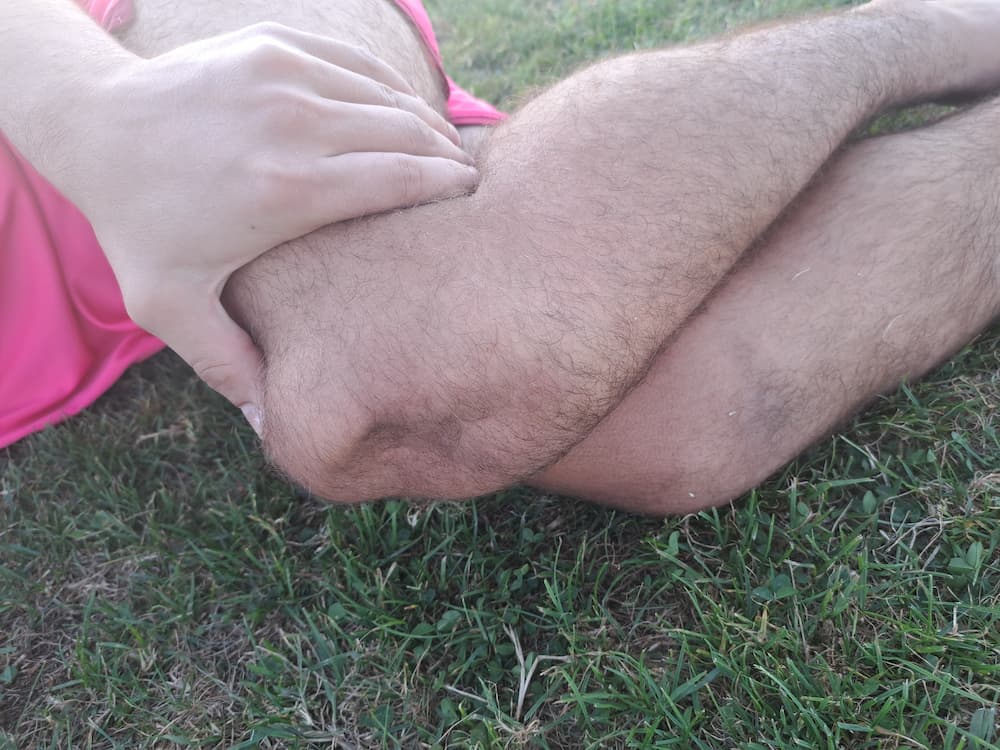
What is the patella?
The patella (kneecap) is the largest sesamoid bone in the human body. It is triangular in shape and slightly convex on its front surface, while its back, articular surface is divided by a longitudinal central ridge. The back of the patella carries the thickest cartilage in the human body, reaching up to 7 mm in places. The patella sits at the front of the knee, within a bony groove at the lower end of the femur, and forms part of the knee's extensor mechanism through its connections to the quadriceps tendon at its base (upper pole) and the patellar ligament at its tip (lower pole). The back of the patella is divided by a vertical ridge that matches the groove at the lower end of the femur, preventing excessive sideways movement of the patella. Ligaments forming several layers of the knee joint capsule attach to the sides of the patella and, like guy-ropes, contribute to its stability. The main function of the patella is to increase the distance between the quadriceps muscle and the knee's centre of rotation, lengthening the quadriceps lever arm and boosting the force of knee extension by up to 50%. Together with the femur, the patella forms the patellofemoral joint, an integral part of the knee. Because the patellofemoral joint is shallow and its bony surfaces are not well matched, its stability depends heavily on the soft tissues that surround and stabilise it, both statically and dynamically. For this reason, given its specific bony anatomy and the numerous ligamentous structures and muscles that act dynamically on the patella, the patellofemoral joint is considered, from a biomechanical standpoint, one of the most complex joints in the human body.
What is a primary patella dislocation – traumatic patella luxation in children and adolescents?
Knee injuries are among the most common injuries of the musculoskeletal system in recreational and competitive athletes, but they also occur during everyday activities. In children, knee injuries are even more frequent, accounting for around 5% of all injuries that bring children aged 1 to 18 to the emergency department.
An acute primary patella dislocation (a first-time dislocation of the kneecap) is a traumatic disruption of the patella's normal, previously undisturbed position relative to the femur, usually a shift of the patella towards the outer side of the knee. After ACL rupture, primary patella dislocation is the second most common cause of traumatic bleeding into the knee (haemarthrosis) and accounts for roughly 3% of all traumatic knee injuries. In children under 14, primary patella dislocation is the most common serious knee injury accompanied by acute haemarthrosis. Several studies have shown that female sex and younger age are associated with a higher risk of primary patella dislocation, and that the risk decreases with age. Certain anatomical variations in children – in the shape of the patella, the lower femur, and the alignment of the lower limb axes – also predispose to traumatic patella dislocation. Predisposing factors for increased patellar instability include: dysplasia of the lower femur, a high-riding patella, increased femoral anteversion, increased external tibial rotation, lateral patellar tilt, hypoplasia of part of the quadriceps, subtalar pronation, valgus alignment of the lower leg, and increased ligament laxity.
Acute primary patella dislocation most often occurs due to a knee injury during sport or another physical activity. What is surprising at first glance, however, is that traumatic patella dislocation is usually a non-contact injury. In around 90% of cases, the child was not in physical contact with another person at the moment of injury. The typical mechanism occurs with the knee minimally bent, the lower leg turned outward, and the trunk beginning to rotate. Other, less common causes of traumatic patella dislocation are direct blows to the knee.
How is primary traumatic patella dislocation diagnosed in children and adolescents?
Primary traumatic patella dislocation is a dramatic event, and most children describe the injury as the knee “popping out to the side,” followed by a fall. In some injured children the patella spontaneously returns to its correct position as the knee straightens (spontaneous reduction), while in others active reduction is required. On examination by a paediatric orthopaedic specialist, clinical presentation ranges from extreme pain and marked knee swelling to an almost entirely pain-free knee. Most children limp during the examination and have limited knee movement. Palpation of the knee typically reveals fluid in the joint – haemarthrosis – along with tenderness on the inner side of the patella and the outer side of the femur. If the patella is not excessively painful, the so-called patellar apprehension test can be performed, which is positive if the child fears the patella will “pop out” again. An X-ray of the knee should always be taken in three views: AP (anterior-posterior; front view), LL (latero-lateral; side view), and an axial (skyline) view of the patella. The X-rays are examined for an osteochondral fracture or a loose fragment within the joint, which often forms when a piece of the patella or femur breaks off during the dislocation. If the mechanism of injury is unclear, or a primary traumatic patella dislocation is suspected but clinical tests are inconclusive, an MRI scan is recommended.
In more than 90% of patella dislocations, part of the inner side of the knee joint capsule tears – a ligament called the medial patellofemoral ligament (MPFL) – which can be seen on MRI scans. Since the MPFL is the primary passive stabiliser of the patella, preventing excessive outward movement, its injury can lead to patellar instability. In other words, after a primary traumatic patella dislocation, the chances of a repeat dislocation increase, which is why appropriate treatment is needed. Other knee injuries that occur less frequently alongside patella dislocation include cruciate ligament tears and meniscus damage.
How is primary traumatic patella dislocation treated in children and adolescents?
Every child who has had a primary traumatic patella dislocation should be examined by a paediatric orthopaedic specialist. X-rays allow us to determine whether an osteochondral fracture is present. If a loose osteochondral fragment is found in the knee, urgent surgery is needed to repair the fracture with special screws or, for smaller fragments that cannot be stabilised with screws, to remove the fragment from the knee. The surgical technique generally used is knee arthroscopy, in which small skin incisions allow a camera and appropriate instruments to enter the knee joint to perform the procedure. Occasionally, a larger incision on the knee is needed to fix the bone fragment with screws. During surgery it is also good practice to repair (suture) the inner patellar ligaments to make the patella more stable.
If no osteochondral injuries are seen on X-ray, there is no indication for emergency surgery, and the knee is best immobilised in a brace or plaster splint with the knee straight for a few days. During immobilisation the knee must not be bent, giving the capsular ligaments and the MPFL a chance to heal. While wearing the immobiliser, especially in the first days after the injury, cold compresses should be applied to the knee, and physical therapy to maintain quadriceps muscle tone should be carried out throughout. In most cases of patella dislocation, once immobilised children can walk freely, bearing weight on the injured leg up to the point of pain, without needing to be completely non-weight-bearing on crutches. A week after the injury, a patellar brace with a round front opening is fitted to the knee, and children gradually begin active physical therapy.
How are recurrent patella dislocations – patellar instability – treated in children and adolescents?
In most injured children, after treatment of a first traumatic patella dislocation with immobilisation and physical therapy, no instability will develop, while recurrent dislocation occurs in 15% to 44% of cases. If repeated patella dislocations occur and the child has a feeling of knee instability despite adequate conservative treatment with physical therapy, a new clinical examination by a paediatric orthopaedic specialist is needed. During the examination, particular attention is paid to sideways mobility of the patella; a shift of more than 50% of the patella's width to the side is considered excessive and typically causes a feeling of apprehension about further dislocation. The examination also considers other factors contributing to patellar instability, such as leg alignment, ligament laxity, foot position and quadriceps muscle strength.
Recurrent instability – repeated patella dislocation – occurs more frequently in children and can cause damage to the joint cartilage, osteochondral fractures, permanent patellar instability, pain, reduced activity, and consequent degenerative changes in the patellofemoral joint. When the patella is unstable and children feel limited in everyday activities, surgery is needed. MPFL reconstruction is often indicated to stabilise the patella. During reconstruction, tendons taken from around the same knee being operated on are used to replace the original, native MPFL. MPFL reconstruction may be performed as a stand-alone procedure or as part of a more complex knee procedure, depending on the findings of further work-up. After MPFL reconstruction, the knee is typically immobilised in a brace for a few weeks, and physical therapy is carried out according to appropriate protocols. Return to sporting activities is expected after 4–5 months. Treatment of patellar instability with MPFL reconstruction has proven successful – the patella becomes more stable, and children can return to the activities that fulfil and delight them.
For more information or to schedule an appointment, please get in touch via the contact form.
Book Appointment