What is scoliosis?
Scoliosis is a deviation of the spine to the side by more than 10o, leading to the formation of an unnatural curve in the spine. It can be structural – true scoliosis, which is essentially a spinal disease, and postural scoliosis, which arises as a result of altered biomechanical relationships, such as leg length discrepancies or pelvic tilt.
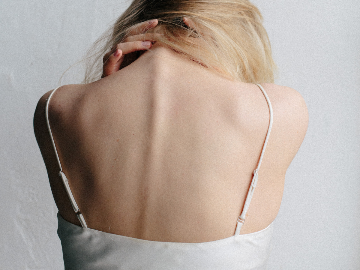
Structural scoliosis, based on the age of onset, can be classified into early-onset scoliosis, occurring in children up to ten years of age, and adolescent scoliosis. Adolescent idiopathic scoliosis is by far the most common and typically occurs in girls with the appearance of right-sided thoracic curvature. Since structural scoliosis involves three-dimensional deformations, lateral curvature is always accompanied by vertebral rotation and torsion of the entire spine. In children, a characteristic hump – gibbus can be observed on the back. The cause of adolescent idiopathic scoliosis is unknown, but genetics has been shown to have the most significant influence. The essential factor for the development of adolescent idiopathic scoliosis is the uneven and uncoordinated growth of the vertebral body’s front and back parts. Specifically, the front part of the vertebra (vertebral body) grows faster than its back part (vertebral arches), leading to their rotation. Other factors mentioned as possible causes of adolescent idiopathic scoliosis include premature growth spurt, reduced bone mineral density – osteoporosis, significant leg length discrepancy, and the development of muscles along the spine. Parents often mention that their children sit or stand hunched over or engage in asymmetrical sports, attributing these factors to the development of scoliosis. However, these circumstances do not affect the onset of structural scoliosis.
How to diagnose scoliosis?
Children and parents usually come for spine examinations after noticing poor posture, a protrusion on the back, or shoulder height asymmetry, or due to back pain. Sometimes, a child may not have any of the mentioned symptoms but is referred to a pediatric orthopedist after a school systematic examination. The majority of children coming for spine examinations subjectively do not have any difficulties, but if there is pain, it is crucial to thoroughly examine the character of the pain, its duration, and its impact on the appearance of the spine.
For instance, suddenly occurring back pain causing twisted posture can be a sign of inflammation, tumor diseases, intervertebral disc injury, or spondylolysis (stress fracture of the vertebral arch). For girls, it is important to know the time of the first menstruation (menarche) because it is believed that the growth of the spine is most pronounced a year before and two years after menarche, and it is during this period of the most intensive spinal growth that the largest deformities occur.
During the clinical examination of the spine, the most important aspect is performing the forward bending test, observing the contours of the spine, and looking for asymmetry. Typically, in idiopathic adolescent scoliosis, a larger protrusion (gibbus) is noticed on the right thoracic side and a smaller protrusion on the left lumbar side as muscle asymmetry. Clinically, the height of the pelvic wing bones is generally symmetrical. If there is significant asymmetry in the height of the pelvic wing bones, suspicion of leg length discrepancy arises, and wooden blocks should be placed under the shorter leg, followed by a re-examination of the spine in forward bending. It is considered that a difference in leg length of up to two cm does not affect the occurrence of structural scoliosis. However, due to the difference in leg length, pelvic tilt and consequently postural scoliosis can easily occur, which can be easily corrected.
During each spine examination, attention should be paid to the front of the chest, observing the relationships of rib arches with the breastbone. Sometimes pectus carinatum (chicken chest) or, conversely, pectus excavatum (hollowed chest) may be observed. Children should walk on their toes and heels to rule out neurological deficits. The basic radiological imaging of the spine in cases of suspected scoliosis is an X-ray of the spine in the posterior-anterior (PA) projection. Children must stand on their feet and be barefoot during imaging, and it is ideal if the same X-ray image shows the spine and the pelvic wing bones, Figure 1. If the X-ray shows a lateral curvature of the spine of 11o Cobb or more, the child has scoliosis. According to orthopedic criteria, up to 10o lateral curvature of the spine is not scoliosis in the full sense of the word. In the same X-ray image, attention should be paid to the bone maturity of the pelvic wing bones, which is extremely important in choosing the right therapy. As mentioned earlier, painful scoliosis that is also progressive can be a sign of other diseases that require urgent treatment. Therefore, children with progressive painful scoliosis should be urgently referred for MRI to better visualize the structure of the vertebrae and soft tissues around the spine.

How to treat scoliosis?
The most crucial prognostic factors for adolescent idiopathic scoliosis are:
- Cobb angle
- Age of the child
- Skeletal maturity
- For girls, the onset of the first menstruation
It is believed that younger age and a larger Cobb angle at the diagnosis of adolescent idiopathic scoliosis influence its progression. In about 80% of cases, scoliosis worsens when the Cobb angle exceeds 25o of lateral curvature.
There are three main treatment goals for adolescent idiopathic scoliosis: to halt its progression, correct the spinal deformity, and maintain the corrected spinal shape. Every child with scoliosis should undergo specific physical therapy. Sports activities, preferably swimming, are recommended. However, it is essential to involve the child in a sport they are motivated to participate in. Since adolescence is a sensitive period for young individuals’ development, imposing unwanted activities excessively may have a negative impact on their engagement in sports.
If, at the discovery of adolescent idiopathic scoliosis, the Cobb angle measures more than 20o of lateral curvature, and skeletal maturity is not complete (i.e., the child still has growth potential), spinal brace should be included in the treatment. The brace is applied based on the three-point system and corrects the spine by directing the growth forces in the corrected direction. The main task of the brace is to stop scoliosis progression, and in some cases, it can achieve up to a 20% correction of the scoliotic deformity. When prescribing the brace, it is crucial to have a careful discussion with the children since they are sensitive during this period of life and may find it aesthetically displeasing. Therefore, it is necessary to explain the principles of scoliosis treatment with a brace in detail, emphasizing that the brace should be worn for at least 16 hours daily.
Children undergoing scoliosis treatment should be clinically monitored and undergo spinal X-ray imaging at least once a year. If there is worsening of scoliotic curvature beyond 40o in the thoracic spine or 50o in the lumbar spine, surgical intervention should be considered. Surgery involves stopping the spinal growth and correcting the scoliotic deformity through spinal fusion techniques.
A much rarer type of scoliosis, early-onset scoliosis (occurring before the age of ten), requires active treatment immediately after diagnosis. Corrections with casting may be performed, gradually replaced by a plastic brace. The general idea is to achieve maximum spinal growth in the correct direction before surgery since surgery essentially stops spinal growth by connecting the vertebrae, leading to spinal stiffness. Unfortunately, this type of scoliosis always requires surgical intervention as it progresses rapidly and compromises breathing.
Postural scoliosis is treated by correcting statics and addressing leg length discrepancies, usually with orthopedic insoles for both feet, with the thicker one placed under the shorter leg to functionally lengthen it. If the leg length difference exceeds two cm, corrective orthopedic shoes may be required. Leg length differences can also be treated surgically: if children are still growing, the growth of the longer leg can be slowed or stopped; after growth is complete, the shorter leg can be lengthened. Alongside static correction, physical therapy, including stretching exercises for the muscles around the pelvis and thighs, is essential.